Multi-Modal AI for Medical Specialists
Accelerating diagnosis across Radiology, Pathology, Oncology, and Cardiology. AI-powered analysis in minutes, not hours.
Pathology
Tissue & Biopsy Analysis
Radiology
CT, MRI & X-ray Scans
Oncology
Tumor Detection & Analysis
Cardiology
Cardiac Imaging & ECG
Reduce Diagnosis Time
From hours to minutes
Multi-Modal Analysis
All imaging types, one platform
Scales With Your Growth
From 10 to 10,000+ cases/day
Understanding OneMedAI's Approach
A 2-minute overview of how our multi-modal AI platform assists medical specialists in delivering faster, more accurate diagnoses across multiple departments.
3 Minutes
Complete Overview
Multi-Modal AI
All Imaging Types
Specialist-Focused
Augmentation, Not Replacement
What You'll Get
How OneMedAI processes multiple medical imaging modalities
Why our AI reduces diagnosis time from hours to minutes
Our approach to data security and regulatory compliance
How specialists maintain full control and oversight
Real-world results from early partner hospitals
Scalability from 10 to 10,000+ cases per day
Ready to see OneMedAI in your workflow?
See What OneMedAI Sees
Real-time AI analysis of a brain MRI scan showing how OneMedAI identifies, analyzes, and reports findings with radiologist-level precision.
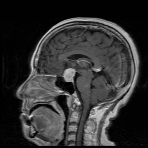
Primary Findings
Sellar/Suprasellar Mass
- Pituitary macroadenoma (most likely)
- Size: 15-20mm craniocaudal
- Suprasellar extension present
- Compressing optic chiasm
Mass Effect Detected
- Superior displacement of optic chiasm
- Risk of visual field deficits
- Immediate evaluation recommended
Surrounding Structures
- No hydrocephalus detected
- No herniation present
- Brainstem unremarkable
- Ventricles within normal limits
Differential Diagnosis
AI Recommendations
Imaging
Dedicated pituitary MRI protocol with contrast
Laboratory
Endocrine panel: Prolactin, IGF-1, cortisol, TSH
Ophthalmology
Visual field testing and acuity assessment
Referrals
Endocrinology and neurosurgery consults
AI-Powered Solutions Across All Specialties
From diagnostic imaging to specialized analysis, OneMedAI delivers comprehensive AI assistance across every major medical department.
Pathology
Tissue & Biopsy Analysis
Radiology
CT, MRI & X-ray Scans
Oncology
Tumor Detection & Analysis
Cardiology
Cardiac Imaging & ECG
Dermatology
Skin Lesion Analysis
Neurology
Brain Imaging & Diagnostics
Ophthalmology
Retinal Imaging & Eye Scans
Pulmonology
Chest X-rays & Lung Scans
Gastroenterology
Endoscopy & GI Imaging
Orthopedics
Bone & Joint Imaging
Hematology
Blood Analysis & Microscopy
Nephrology
Kidney & Urinary Imaging
Your Expertise + Our AI = Better Outcomes
OneMedAI handles routine analysis so specialists can focus on complex cases and patient care. We augment human expertise, never replace it.
Specialist Alone
Traditional WorkflowManual Image Review
15-20 min per case
Pattern Recognition
Fatigue after 50+ cases
Report Writing
10-15 min documentation
Final Review
Limited time for complex cases
Specialist + OneMedAI
AI-AugmentedAI Pre-Analysis
2-3 min automated scan
AI Highlights Anomalies
ROI markers + confidence scores
Auto-Generated Draft
Structured report ready
Specialist Focuses on Complex Cases
More time for critical thinking
Meet the team that makes the magic happen
Meet our diverse team of world-class creators, designers, and problem solvers.

Santhosh P.
Founder & CEO

Mandar J.
CMO

Vinod B.
Chief Innovation Officer

Nagalakshmi V.
Head of Operations

Suresh Iyer
Chief Product Owner

Yasir Noman
Chief Advisor

Vilas Junagade
Chief Financial Advisor
Development Team
Akanksha Kalaskar
AI Developer
Zeshan Ahmad
Senior Full-Stack Developer
Moiz Ansari
Senior Frontend Developer
Ahmad Ali
Backend Developer
Real-World AI Diagnostic Success
Explore how OneMedAI identifies critical findings across diverse pathologies. Each case study demonstrates our AI's clinical precision and diagnostic capabilities.
Pituitary Macroadenoma with Chiasmal Compression
AI successfully identified a sellar/suprasellar mass with superior displacement of the optic chiasm, demonstrating 95% confidence in detecting urgent findings requiring immediate neurosurgical evaluation.
Early-Stage Lung Nodule Detection
OneMedAI detected a 6mm ground-glass opacity in the right upper lobe that was initially overlooked in preliminary review. Follow-up confirmed early-stage adenocarcinoma, enabling timely intervention.
Parathyroid Adenoma Detection with Chief Cell Pattern
AI successfully identified a well-circumscribed parathyroid lesion with characteristic chief cell predominance, trabecular architecture, and marked reduction in adipose tissue. Analysis detected subtle features distinguishing adenoma from hyperplasia, recommending confirmatory PTH immunohistochemistry and clinical correlation with serum calcium levels.
Normal Sinus Rhythm with Subtle Early Repolarization
AI analysis identified normal sinus rhythm with regular RR intervals, normal axis, and appropriate QRS morphology across all 12 leads. Subtle early repolarization pattern detected in precordial leads V4-V6, classified as benign variant. No ST-segment elevation, depression, or T-wave abnormalities requiring intervention.
Chronic Bilateral Pars Defects with Spondylolisthesis
AI detected bilateral pars interarticularis defects at L5 with sclerotic margins indicating chronicity, and identified associated Grade I anterolisthesis of L5 on S1. Analysis highlighted critical features distinguishing chronic spondylolysis from acute fracture, enabling appropriate conservative management planning.
Malignant Melanoma Detection with ABCDE Criteria
AI analysis identified a pigmented lesion exhibiting asymmetry, irregular borders, color variation (brown, black, pink tones), diameter >6mm, and evolving characteristics. Dermoscopic features including irregular pigment network and blue-white veil prompted urgent biopsy recommendation. Histopathology confirmed superficial spreading melanoma, demonstrating AI's capability in early melanoma detection.
Want to see how OneMedAI performs on your cases?
Transform Your Medical Practice Today
Choose the solution that fits your needs and experience the future of AI-powered diagnostics
Enterprise Solution
For Hospitals & Medical Centers
Full-scale AI integration for your entire radiology department with dedicated support
- Custom implementation plan
- Dedicated account manager
- Staff training & onboarding
- 24/7 priority support
Individual Practice
For Independent Specialists
Perfect for solo practitioners and small clinics looking to enhance diagnostic accuracy
- Flexible pricing plans
- Quick setup in 48 hours
- No long-term contracts
- 30-day money-back guarantee